


 Foundational characteristics of cancer include proliferation, angiogenesis, migration, evasion of apoptosis, and cellular immortality. Find key markers for these cellular processes and antibodies to detect them.
 The SUMOplot™ Analysis Program predicts and scores sumoylation sites in your protein. SUMOylation is a post-translational modification involved in various cellular processes, such as nuclear-cytosolic transport, transcriptional regulation, apoptosis, protein stability, response to stress, and progression through the cell cycle.
 The Autophagy Receptor Motif Plotter predicts and scores autophagy receptor binding sites in your protein. Identifying proteins connected to this pathway is critical to understanding the role of autophagy in physiological as well as pathological processes such as development, differentiation, neurodegenerative diseases, stress, infection, and cancer.
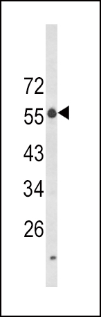
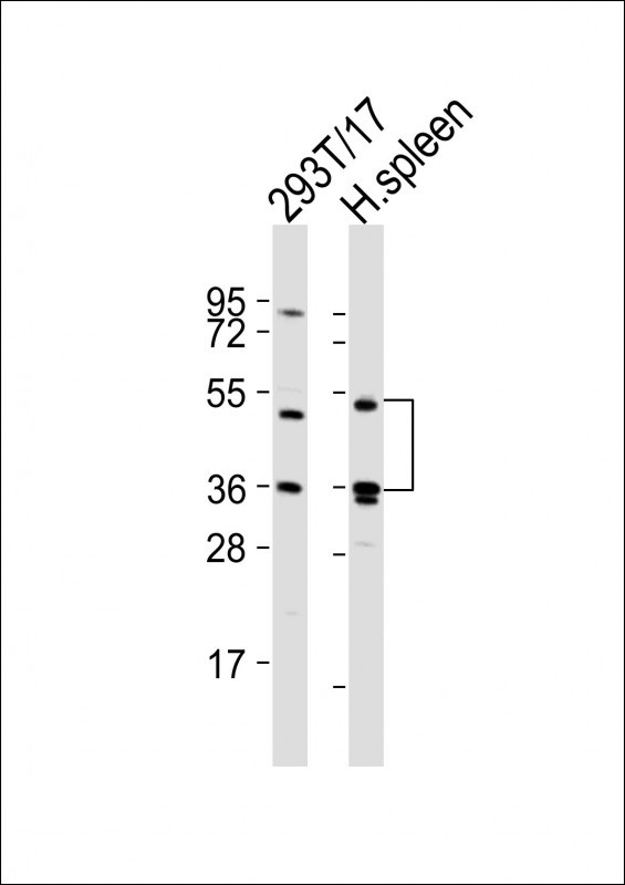
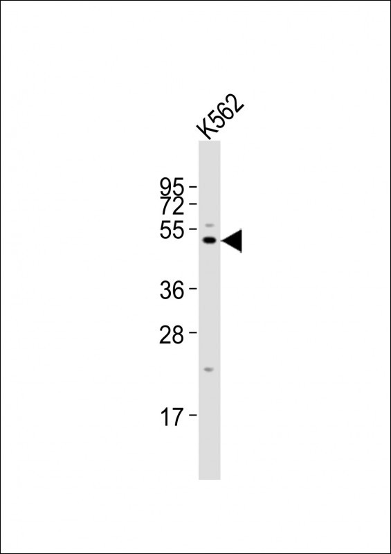
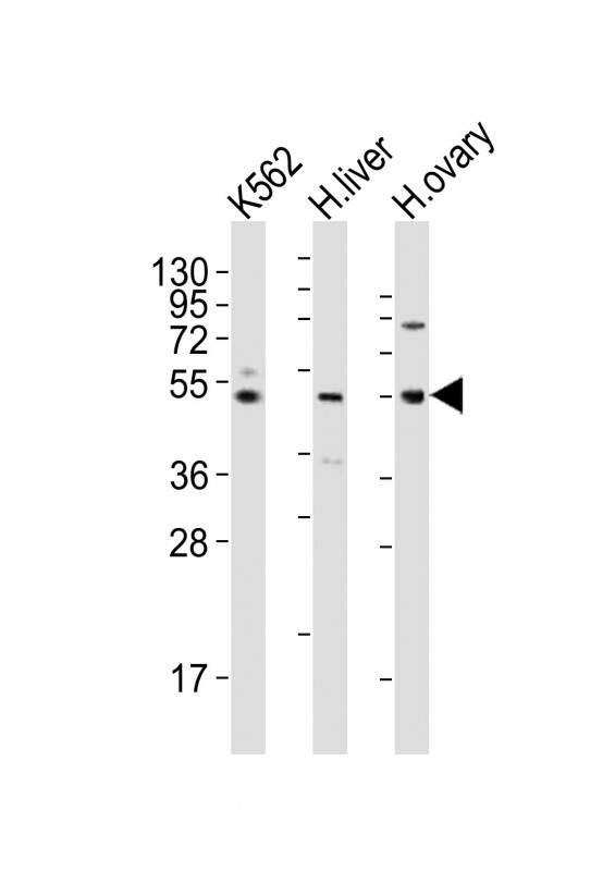
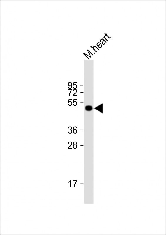
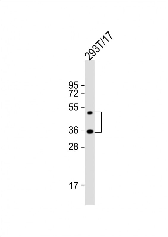
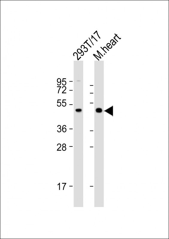
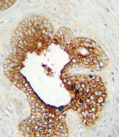
LAG3 Antibody (Center)
Affinity Purified Rabbit Polyclonal Antibody (Pab)


- SPECIFICATION
- CITATIONS
- PROTOCOLS
- BACKGROUND

Application
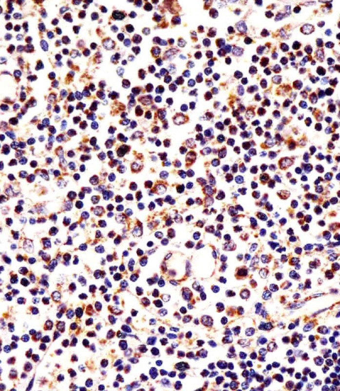
| WB, IHC-P, FC, E |
|---|---|
| Primary Accession | P18627 |
| Reactivity | Human, Mouse |
| Host | Rabbit |
| Clonality | Polyclonal |
| Isotype | Rabbit IgG |
| Calculated MW | 57449 Da |
| Antigen Region | 103-132 aa |
| Gene ID | 3902 |
|---|---|
| Other Names | Lymphocyte activation gene 3 protein, LAG-3, Protein FDC, CD223, LAG3, FDC |
| Target/Specificity | This LAG3 antibody is generated from rabbits immunized with a KLH conjugated synthetic peptide between 103-132 amino acids from the Central region of human LAG3. |
| Dilution | WB~~1:1000 IHC-P~~1:25 FC~~1:25 E~~Use at an assay dependent concentration. |
| Format | Purified polyclonal antibody supplied in PBS with 0.09% (W/V) sodium azide. This antibody is purified through a protein A column, followed by peptide affinity purification. |
| Storage | Maintain refrigerated at 2-8°C for up to 2 weeks. For long term storage store at -20°C in small aliquots to prevent freeze-thaw cycles. |
| Precautions | LAG3 Antibody (Center) is for research use only and not for use in diagnostic or therapeutic procedures. |
| Name | LAG3 {ECO:0000303|PubMed:35761082, ECO:0000312|HGNC:HGNC:6476} |
|---|---|
| Function | [Lymphocyte activation gene 3 protein]: Inhibitory receptor on antigen activated T-cells (PubMed:20421648, PubMed:35761082, PubMed:7805750, PubMed:8647185). Delivers inhibitory signals upon binding to ligands, such as MHC class II, its main ligand present at the surface of antigen-presenting cells (APCs), and FGL1, which is secreted by hepatocytes and certain types of tumor cells (PubMed:30580966, PubMed:32920841, PubMed:35761082, PubMed:39671469, PubMed:7589152, PubMed:8647185, PubMed:9159144). Ligand-binding initiates a signaling that inhibits the T-cell receptor (TCR) in the immunological synapse, preventing T-cell activation (PubMed:40101708). Mechanistically, ligand-binding promotes (1) ubiquitination of the KIEELE motif, unleashing the RRFSALE motif from the membrane and (2) leading to the formation of condensates with the TCR component CD3E, thereby disrupting the association between CD3E and LCK and preventing TCR activation (PubMed:40101708, PubMed:40592325). May inhibit antigen- specific T-cell activation in synergy with PDCD1/PD-1 (By similarity). Negatively regulates the proliferation, activation, effector function and homeostasis of both CD8(+) and CD4(+) T-cells (PubMed:20421648, PubMed:7805750, PubMed:8647185). Also mediates immune tolerance: constitutively expressed on a subset of regulatory T-cells (Tregs) and contributes to their suppressive function (By similarity). Also acts as a negative regulator of plasmacytoid dendritic cell (pDCs) activation (By similarity). |
| Cellular Location | [Lymphocyte activation gene 3 protein]: Cell membrane; Single-pass type I membrane protein. Note=Clusters on the T-cell surface following ligand-binding |
| Tissue Location | Primarily expressed in activated T-cells and a subset of natural killer (NK) cells. |

Thousands of laboratories across the world have published research that depended on the performance of antibodies from Abcepta to advance their research. Check out links to articles that cite our products in major peer-reviewed journals, organized by research category.
info@abcepta.com, and receive a free "I Love Antibodies" mug.

Provided below are standard protocols that you may find useful for product applications.
Background
Lymphocyte-activation protein 3 belongs to Ig superfamily and contains 4 extracellular Ig-like domains.
References
Smyth,D.J., et.al., BMC Med. Genet. 7, 20 (2006)
If you have used an Abcepta product and would like to share how it has performed, please click on the "Submit Review" button and provide the requested information. Our staff will examine and post your review and contact you if needed.
If you have any additional inquiries please email technical services at tech@abcepta.com.
Ordering Information
Other Products
Shipping Information
